All images / illustration
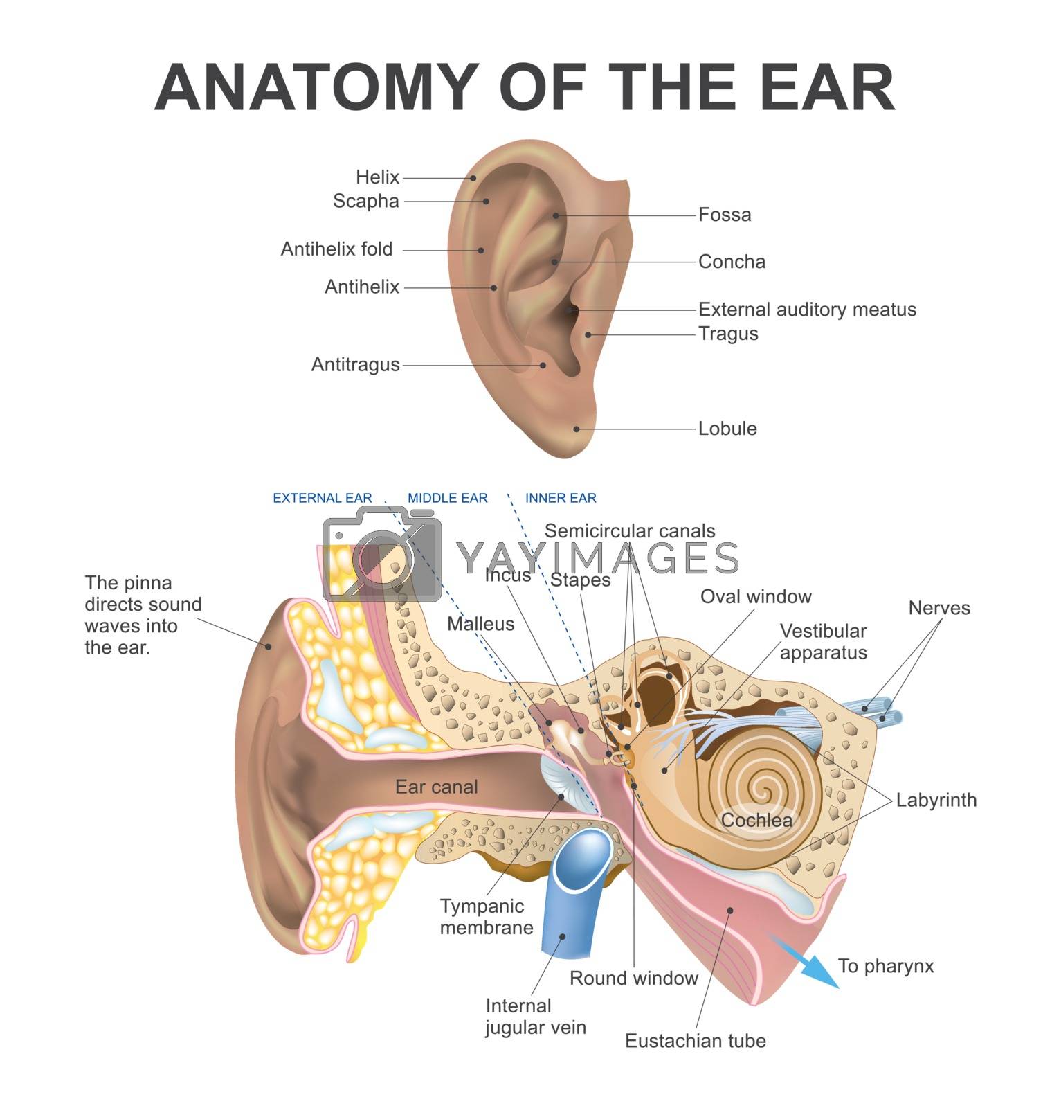
Anatomy of the ear.
The human ear consists of three parts the outer ear, middle ear and inner ear. The ear canal of the outer ear is separated from the air filled tympanic cavity of the middle ear by the eardrum. The middle ear contains the three small bones the ossicles involved in the transmission of sound, and is connected to the throat at the nasopharynx, via the pharyngeal opening of the Eustachian tube. The inner ear contains the otolith organs the utricle and saccule and the semicircular canals belonging to the vestibular system, as well as the cochlea of the auditory system.
-
License:
Royalty Free License.
The license type determines how you can use this image.

Std. Ext. Print / Editorial Graphic Design Web Design Social Media Edit & Modify Resale Items 1 Unlimited Runs - Please see licensing information by clicking here
Vector
All images / illustration
Size Selection:
You can re-download any size after the purchase.
Keywords
-
acoustic
anatomical
anatomy
antihelix fold
auditory
balance
biology
body
canal
cavity
cochlea
cochlear
cross
drawing
ear
eardrum
eustachian
external
health
hear
helix
human
human body
illustration
inner
inner ear
internal
males
medical
membrane
middle
nerve
organ
outer
part
pinna
science
section
semicircular canals
sense
sensory
sound
stapes
Tagus
tube
tympanic
tympanic membrane
vector
vestibular
vestibular apparatus